What Are Bone Preservation Implants and How Do They Work?
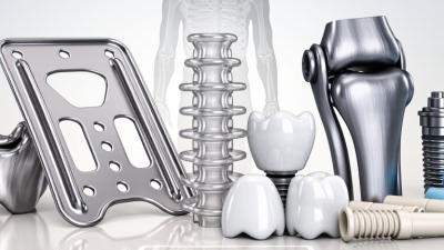
Bone preservation implants are designed to support missing teeth while helping maintain the natural shape and volume of surrounding jawbone. They are not simply smaller or newer dental implants. Their performance depends on implant geometry, surface design, surgical technique, loading forces, and the patient’s healing response. In practice, preservation begins before placement. A clinician may examine a three-dimensional scan, assess bone thickness, and plan an implant position that protects the facial bone wall. Small details matter, including the implant’s thread pattern and its connection to the final crown.
The biology is central. After tooth loss, the jawbone often shrinks because it no longer receives normal stimulation. Bone preservation implants aim to distribute chewing forces more favorably and encourage stable integration with living bone. Per-Ingvar Brånemark, the pioneering Swedish implant researcher, described osseointegration as “a direct structural and functional connection between ordered, living bone and the surface of a load-carrying implant.” That principle still guides modern implant treatment.
However, the phrase can sound more certain than the evidence allows. No implant can guarantee complete bone preservation. Surgical trauma, infection, smoking, poor hygiene, uncontrolled diabetes, and excessive loading may still contribute to bone loss. Results vary.
A careful explanation should separate marketing claims from measurable outcomes. This article will examine how these implants work, which design features may support bone stability, and why long-term maintenance remains essential. The patient’s anatomy always matters. Expect nuance, not promises.
Definition and Purpose of Bone Preservation Implants
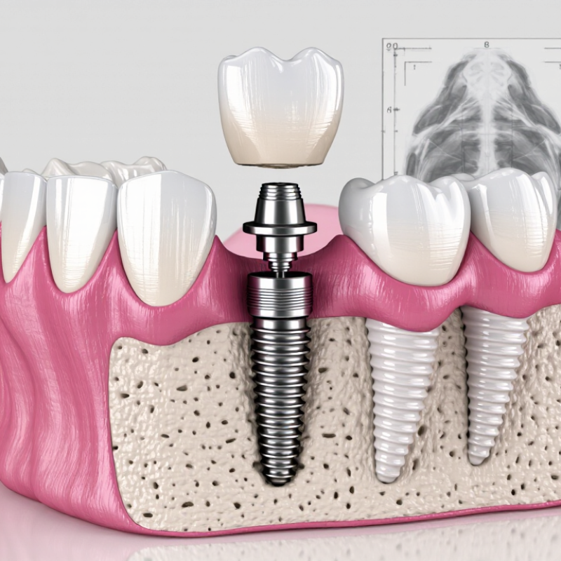
Bone preservation implants are dental implants designed to replace a missing tooth while helping maintain the jawbone around the treatment site. After tooth loss, the bone no longer receives normal chewing pressure. Over time, it may shrink in height and width. An implant transfers controlled biting forces into the bone, which can support healthier bone maintenance.
The purpose is both functional and structural. The implant supports a crown, bridge, or denture, while its root-like shape occupies the space left by the natural tooth root.
Proper placement depends on three-dimensional imaging, bone volume, gum health, and bite forces. In some cases, a dentist may recommend bone grafting before or during implant treatment. An implant cannot create missing bone by itself.
The term “bone preservation” can sound more certain than it really is. Bone changes vary between patients. Infection, smoking, poor cleaning, and excessive pressure may reduce long-term stability. Careful planning matters. Experienced clinicians assess healing conditions, medical history, and daily habits before choosing an approach. Regular reviews and gentle cleaning around the implant also protect the surrounding tissues. This is not a quick fix. It is a biological treatment that requires time, maintenance, and realistic expectations.
How Bone Preservation Implants Work
Bone preservation implants are designed to replace a missing tooth while helping protect the surrounding jawbone. After extraction, the empty socket can shrink because it no longer receives normal chewing pressure. An implant fills that space with a stable, biocompatible post. The term can sound precise, but it is not always used consistently. Treatment plans differ between patients.
How Bone Preservation Implants Work
A dentist places the implant into prepared bone, often after reviewing three-dimensional scans. The implant can support a crown and provide controlled stimulation during chewing. This pressure may encourage the bone to remain more functional and less likely to resorb. In some cases, the dentist adds bone graft material or a protective membrane. These materials support the healing area while new bone develops around the implant surface. Healing takes time. It is not instant.
The implant must remain stable during early healing. Poor oral hygiene, smoking, untreated gum disease, or excessive biting forces can interfere with this process. Professional assessment matters because thin bone, infection, and medical conditions may change the approach. I would not describe these implants as a guaranteed shield against bone loss. Biology can be unpredictable, and even careful treatment may need adjustment.
Tips: Ask how much bone is present before surgery. Request a clear healing timeline. Keep the site clean as instructed, and attend every follow-up visit. Report swelling, persistent pain, or movement promptly. Small details often influence long-term stability.
Types and Materials Used in These Implants
What Are Bone Preservation Implants and How Do They Work?
Bone preservation implants help maintain jawbone volume after tooth loss. They usually include an endosseous implant, placed inside the jaw, and sometimes a graft or barrier membrane. The implant transfers chewing forces into the bone. This stimulation may reduce the shrinkage that follows extraction, although results vary between patients.
Titanium remains widely used because it is strong, lightweight, and generally compatible with bone tissue. Its textured surface supports osseointegration, where living bone grows closely around the implant. Zirconia is another option. It is ceramic, metal-free, and tooth-colored, but its clinical use requires careful case selection.
Neither material is perfect. Bone quality, bite pressure, smoking, and daily cleaning can influence stability.
Graft materials differ as well. Autografts use the patient’s own bone and may heal predictably, but they require a second surgical site. Allografts come from screened human donors. Xenografts use processed animal-derived mineral, while alloplasts use synthetic calcium-based materials. Resorbable collagen membranes can hold graft particles in place during healing. Nonresorbable membranes or titanium mesh may provide stronger support, but removal can require another procedure. The decision is rarely simple. Three-dimensional imaging, medical history, and long-term maintenance should guide material selection. Even experienced clinicians may revise the plan when the defect is larger than expected.
Placement Procedure and Healing Process
Bone preservation implants replace missing tooth roots while helping maintain jawbone structure. Placement begins with a clinical examination, dental imaging, and a review of your medical history. Your clinician checks bone volume, gum health, bite, and healing risks. Sometimes, a bone graft is needed before implant placement. That can add several months. Careful planning matters.
During placement, local anesthesia keeps the area numb. Sedation may be discussed when appropriate. The clinician creates a precise channel in the jawbone and inserts the implant body. A temporary cover or healing component protects the site. Stitches may be used. You may notice pressure, swelling, or mild bruising afterward, but sharp or worsening pain needs prompt review. Keep the area clean as instructed, choose soft foods, and avoid smoking because it can reduce blood supply. Not every surgery heals on schedule.
Over the next weeks, bone gradually bonds with the implant through osseointegration. Follow-up visits assess tissue closure, stability, and infection signs. Once healing is adequate, the replacement tooth is attached and checked for comfort and bite. Healing may take longer with diabetes, gum disease, poor bone quality, or certain medicines. Healing is not perfectly predictable. Ask about realistic timelines and warning signs before treatment.
Benefits, Risks, and Clinical Considerations
Bone Preservation Implants and How They Work
Benefits, Risks, and Clinical Considerations
A bone preservation implant replaces a missing tooth while helping maintain jawbone structure. After extraction, bone may shrink because chewing stimulation disappears. A titanium or ceramic implant sits inside the jawbone and gradually bonds with living bone through osseointegration. This process supports a crown and may reduce the need for removable appliances. It does not recreate natural bone completely. That distinction matters.
Clinical benefits may include improved chewing, stable tooth replacement, and better support for nearby tissues. Preserving bone may also help maintain facial contours over time. Yet results depend on bone volume, gum health, bite forces, and daily cleaning. Surgery can cause swelling, bruising, discomfort, infection, nerve irritation, or implant failure. Smoking, uncontrolled diabetes, and certain medications may increase risk. Rarely, an implant can affect nearby anatomical structures.
A careful assessment should include dental imaging, medical history, gum examination, and realistic expectations. Some patients need bone grafting before or during placement. Immediate placement is not suitable for every extraction site, especially with active infection or major bone loss. Healing often takes several months, although timing varies. Patients should report persistent pain, looseness, or drainage promptly. Treatment planning is never perfect; imaging can underestimate anatomy, and healing can surprise experienced clinicians. Long-term maintenance still requires professional reviews and meticulous home care.
Related Posts
-
Top Biocompatible Implant Materials for Global Sourcing
-
China Best Minimally Invasive Arthroplasty Techniques and Innovations?
-
2026 Best Knee Implant Systems for Optimal Joint Replacement Solutions?
-

Top Prosthesis Fixation Methods You Should Know?
-

China Top Orthopedic Quality of Life Improvements for Patients?
-
How to Choose Biocompatible Implant Materials for Medical Applications?